
Electromyography (EMG) is a diagnostic test that allows for the assessment of the health of muscles and the nerve cells that control them. This technique is widely used in neurology and physiatry to investigate disorders affecting muscles and nerves, helping to detect conditions such as neuropathies, myopathies, muscular dystrophies, and other peripheral nervous system disorders. Electromyography is a key procedure for determining the cause of weakness, spasms, and other muscle problems, providing detailed information on neuromuscular function.
What is electromyography?

Electromyography (EMG) is a study technique that measures the electrical activity generated in muscles at rest and during contraction. This examination is performed by inserting a small needle into the muscle, which records the electrical signals that the nerves send to the muscles, providing a detailed view of neuromuscular function.
Electromyography is usually performed together with nerve conduction studies, which measure the speed and intensity of the electrical signals that the nerves send to the muscles. Both tests are complementary and provide a comprehensive analysis of muscle and nerve function.
How is an electromyography performed?
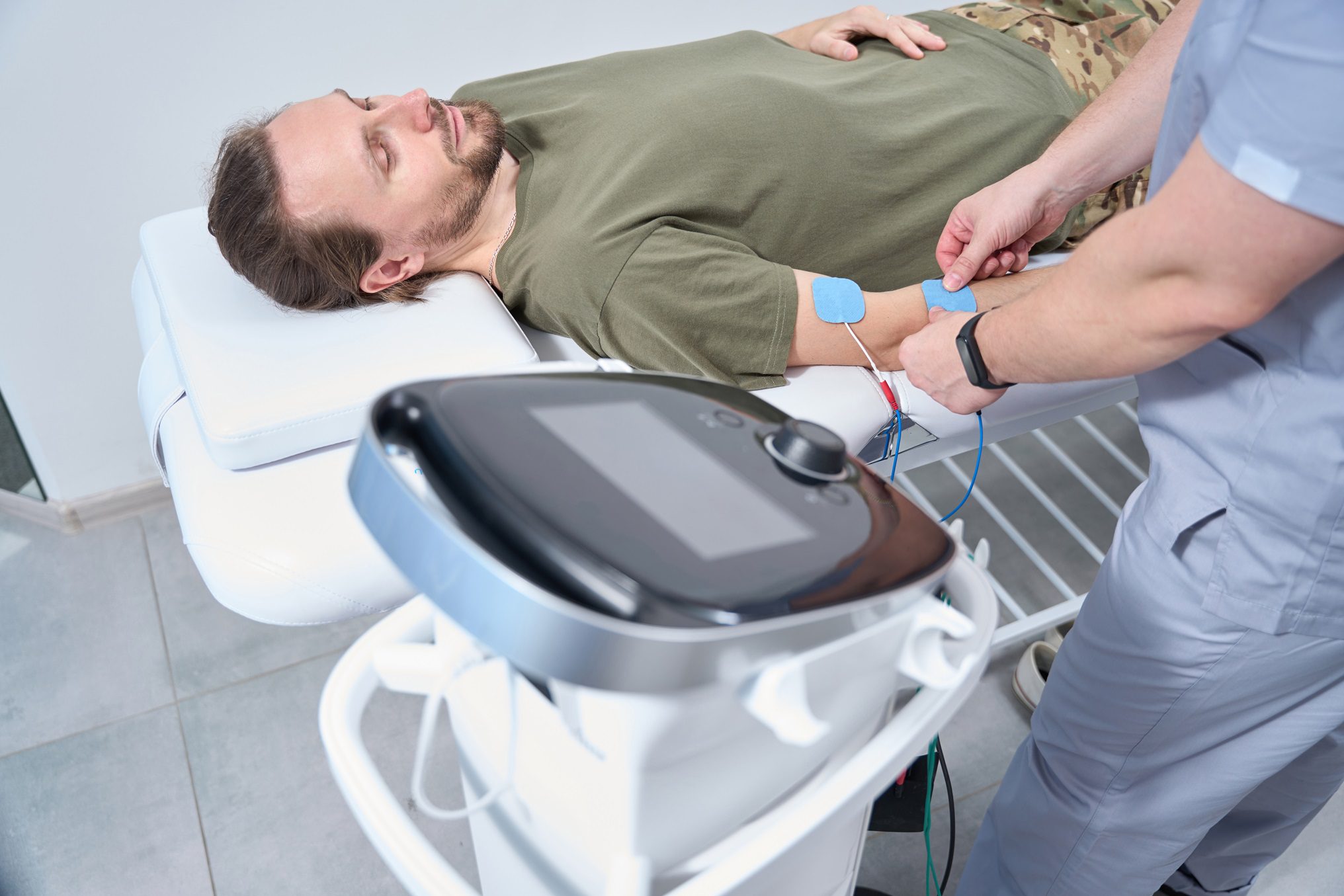
Electromyography is performed in a clinical setting, and the procedure is relatively brief. These are the basic steps of the test:
- Preparation: the physician disinfects the skin in the area of the muscle to be examined. The patient may be instructed to relax and breathe steadily.
- Needle insertion: a thin needle is inserted into the muscle. This needle is an electrode that records the electrical activity of the muscle.
- Measurement at rest and during contraction: the specialist will measure the electrical activity of the muscle while the patient is at rest and will then ask them to perform a slight contraction, such as lifting a leg or flexing an arm.
- Recording of results: the electrical signals are recorded on a monitor, displaying the activity in the form of waves. A neurology specialist will interpret the results to identify any abnormalities in the electrical patterns.
Although the procedure may cause discomfort due to the insertion of the needle, it usually does not require anesthesia, and the discomfort is temporary.
What conditions is electromyography used for?
Electromyography is useful for diagnosing a wide range of neuromuscular disorders. Among the most common conditions for which this study is used are:
- Neuropathies: EMG can help identify damage to the peripheral nerves, whether caused by compression, injuries, or diseases such as carpal tunnel syndrome or diabetic neuropathy.
- Myopathies: muscle diseases, such as muscular dystrophy or myasthenia gravis, can be evaluated through EMG, which allows observation of abnormal patterns in muscle activity.
- Radiculopathy: EMG can detect compression of the nerve roots in the spine, as in cases of herniated disc, which affect the nerves and cause pain or weakness in the limbs.
- Amyotrophic lateral sclerosis (ALS): EMG helps identify the progressive loss of motor neuron function, a characteristic of diseases such as ALS.
- Nerve compression syndromes: conditions such as cubital tunnel syndrome or thoracic outlet syndrome can also be evaluated with electromyography.
Electromyography results and analysis
Los resultados de la EMG se presentan como patrones de ondas que representan la actividad eléctrica del músculo. Un especialista interpretará estos patrones para identificar la posible causa de los síntomas. Algunos de los hallazgos más comunes incluyen:
- Actividad eléctrica en reposo: en condiciones normales, el músculo en reposo debería mostrar poca o ninguna actividad. La actividad en reposo puede indicar daños en los nervios.
- Patrones anormales durante la contracción: un músculo afectado por una enfermedad neuromuscular puede mostrar patrones anormales en la contracción, como señales de baja amplitud o patrones dispersos.
- Espasmos o fasciculaciones: la presencia de espasmos en el registro puede ser un indicador de daño o degeneración en las neuronas motoras, como se ve en condiciones como la ELA.
La interpretación precisa de estos resultados es clave para orientar el diagnóstico y determinar el tratamiento adecuado para la afección específica.
Preparación y cuidados para una electromiografía

Before an electromyography, the physician may suggest some precautions:
- Avoid creams or lotions: patients are advised not to use creams or lotions on the day of the test, as these can interfere with electrode adhesion.
- Inform about medications: some medications, such as anticoagulants, may affect the procedure. It is important for the patient to inform the physician about any medication they are taking.
- Normal diet: fasting is usually not required for electromyography, but it is advisable to avoid caffeine, as it may influence muscle response.
After the test, the patient may experience mild discomfort in the needle insertion area, although this usually disappears within a few hours. If the pain persists or there are signs of infection, the physician should be consulted.
Importance and limitations of electromyography
Electromyography is a valuable tool in the diagnosis of neuromuscular disorders. It provides detailed information on the electrical activity of the muscles and helps physicians identify the source of muscle or nerve problems. However, it also has some limitations:
- Specificity of the condition: EMG can identify whether a muscle or nerve is affected, but further evaluation may be required to determine the exact cause.
- Discomfort during the procedure: for some people, needle insertion may be uncomfortable, although the discomfort is usually mild and temporary.
- Complementary test: in many cases, EMG is only part of the diagnosis, which may require additional tests such as magnetic resonance imaging or laboratory studies.
Despite these limitations, electromyography remains an effective technique for assessing and understanding muscle and nerve problems, guiding appropriate treatment.
Conclusion
Electromyography (EMG) is an essential test for the assessment of muscle and nerve problems. By measuring the electrical activity in the muscles, it enables the diagnosis of conditions such as neuropathies, myopathies, and central nervous system diseases. Although the procedure may cause discomfort, it provides a detailed and accurate view of neuromuscular function.
EMG, together with nerve conduction studies and other diagnostic tests, provides a comprehensive approach to diagnosing neuromuscular conditions, enabling timely and specific treatment. As technology and techniques advance, electromyography will continue to be a cornerstone in the diagnosis and management of nervous system disorders.
If you would like more information about NeuroAiD II, please fill out this contact form.
"*" indicates required fields


