



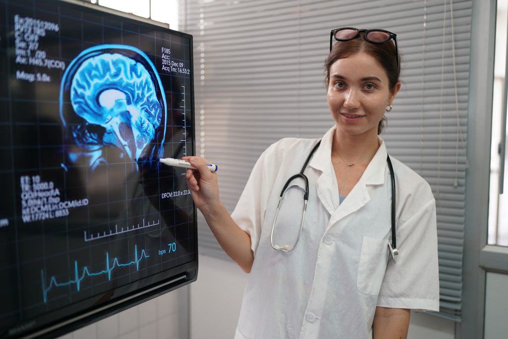
Claude syndrome is a rare neurological syndrome that occurs when a lesion affects a specific region of the midbrain, the upper part of the brainstem. It is characterized by a combination of abnormalities in eye movements and coordination problems on the opposite side of the body, a pattern that helps neurologists precisely localize the brain lesion.
Although it is most commonly associated with stroke, it can also occur as a result of other conditions affecting the brainstem.
What is Claude syndrome?
Claude syndrome is an alternating midbrain syndrome caused by a lesion in the dorsal region of the midbrain.
It is primarily characterized by involvement of:
- The oculomotor nerve (cranial nerve III).
- The red nucleus.
- The superior cerebellar peduncle or its fibers.
The involvement of these structures explains why symptoms occur in one eye and on the opposite side of the body.
Where is the lesion located?
The lesion is typically located in the midbrain tegmentum, a region containing numerous neural pathways involved in motor control and coordination.
This area contains structures responsible for:
- Eye movements.
- Coordination of movement.
- Balance.
- Integration of signals from the cerebellum.
Therefore, a relatively small lesion can produce highly characteristic clinical manifestations.
What is the most common cause?
The most common cause is an ischemic stroke involving small branches of the posterior cerebral artery or the basilar artery.
However, it may also develop as a result of:
- Brainstem hemorrhage.
- Multiple sclerosis.
- Midbrain tumors.
- Vascular malformations.
- Traumatic brain injury.
The nature of the symptoms depends on the size and extent of the lesion.
Symptoms of Claude syndrome
Symptoms have a sudden onset when the underlying cause is vascular.
Third cranial nerve palsy
Involvement of the oculomotor nerve causes symptoms on the same side as the lesion, including:
- Drooping of the eyelid (ptosis).
- Difficulty moving the eye.
- Double vision (diplopia).
- A dilated pupil in some cases.
- Downward and outward deviation of the eye.
These signs are often among the first abnormalities noted during neurological examination.
Coordination disturbances
Damage to the red nucleus and cerebellar pathways causes symptoms on the opposite side of the body, including:
- Ataxia.
- Inaccurate movements.
- Difficulty performing fine motor movements.
- Gait instability.
- Intention tremor.
These disturbances may affect both the contralateral arm and leg.
Other possible symptoms
Depending on the extent of the lesion, other symptoms may also occur:
- Dysarthria.
- Slowness of movement.
- Balance disturbances.
- Difficulty maintaining posture.
- Fatigue while walking.
In most cases, muscle strength remains relatively preserved, which helps differentiate this syndrome from other midbrain syndromes.
How is it diagnosed?
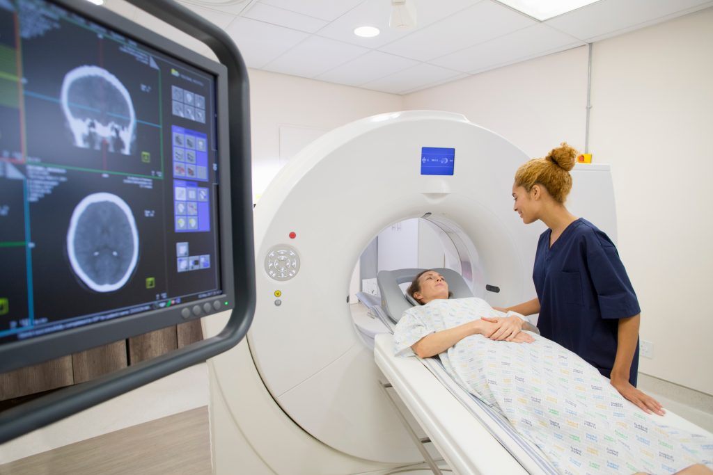
Diagnosis is based on a combination of neurological examination and imaging studies.
Clinical evaluation identifies the association between:
- Ipsilateral third cranial nerve palsy.
- Contralateral ataxia.
- Coordination disturbances.
Subsequent imaging studies may include:
- Brain magnetic resonance imaging (MRI).
- Diffusion-weighted MRI to detect recent infarctions.
- Magnetic resonance angiography (MRA) or computed tomography angiography (CTA).
- Computed tomography (CT) during the acute phase.
In addition, the presence of vascular risk factors and other possible causes of brainstem lesions are usually investigated.
Differences from other midbrain syndromes
Claude syndrome shares some features with other crossed brainstem syndromes, although there are important differences.
These include:
Weber syndrome
It causes:
- Third cranial nerve palsy.
- Contralateral hemiplegia due to involvement of the corticospinal tract.
Benedikt syndrome
It is characterized by:
- Oculomotor nerve palsy.
- Severe tremor.
- Involuntary movements.
- Ataxia.
Nothnagel syndrome
Predominant features include:
- Cerebellar disturbances.
- Ocular palsy.
- Balance disorders.
Identification of the clinical pattern helps localize the lesion with a high degree of precision.
Clinical course and prognosis
Prognosis depends on factors such as:
- The cause of the lesion.
- The size of the infarction.
- How quickly treatment is initiated.
- The patient’s age.
- The presence of other neurological conditions.
In some patients, ocular disturbances improve progressively, while recovery of coordination may require a longer period.
Rehabilitation
Rehabilitation aims to restore the highest possible level of functional ability through an individualized treatment program.
It may include:
- Physiotherapy to improve gait and balance.
- Occupational therapy to restore movement precision.
- Specific coordination exercises.
- Visual rehabilitation when oculomotor disturbances persist.
- Speech and language therapy if speech difficulties are present.
The intensity and duration of the rehabilitation program depend on the residual deficits present in each patient.
Can it be prevented?
When Claude syndrome has a vascular origin, prevention focuses on reducing the risk of stroke by controlling factors such as:
- Arterial hypertension.
- Diabetes.
- High cholesterol.
- Smoking.
- Heart disease.
- Physical inactivity.
Controlling these risk factors also helps reduce the risk of other brainstem lesions.
Conclusion
Claude syndrome is a rare neurological syndrome caused by a lesion in the midbrain, usually resulting from a stroke. The combination of third cranial nerve palsy and coordination disturbances on the opposite side of the body is its most characteristic feature. Recognizing this pattern helps localize the lesion to a highly specific region of the brainstem and facilitates early neurological assessment to determine the most appropriate management.
If you need information about NeuroAiD II, you can fill out this contact form
"*" indicates required fields


