


A basal ganglia infarction is a type of ischemic stroke that affects a group of deep brain structures responsible for regulating movement, motor learning, coordination, and various cognitive functions. Although these regions are not directly involved in generating muscle strength, damage to them can cause significant neurological impairments.
Depending on the size and location of the infarction, symptoms may range from mild weakness to movement disorders or difficulties with coordination.
What is a basal ganglia infarction?

A basal ganglia infarction occurs when blood flow to these deep brain structures is interrupted, causing neuronal damage due to a lack of oxygen.
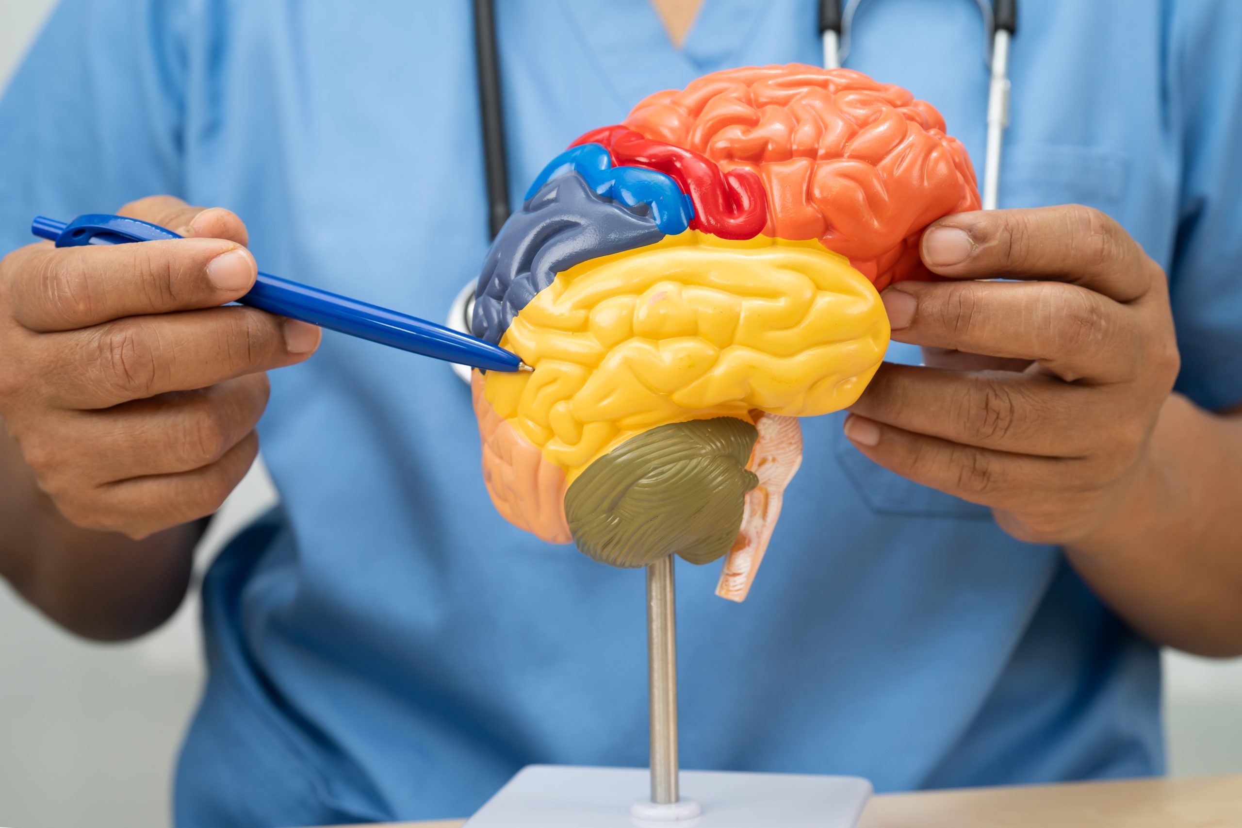
The basal ganglia include several structures, including:
- Caudate nucleus.
- Putamen.
- Globus pallidus.
- Subthalamic nucleus.
- Substantia nigra.
These regions work in coordination with the cerebral cortex and thalamus to regulate the execution of voluntary movements and other brain functions.
What functions do the basal ganglia perform?
The basal ganglia are involved in numerous neurological processes, including:
- Control of voluntary movement.
- Motor coordination.
- Regulation of muscle tone.
- Learning of motor skills.
- Automatization of movements.
- Processing of certain cognitive and emotional functions.
When these structures are damaged, characteristic motor impairments may occur.
Causes
The most common cause is occlusion of the small perforating arteries that supply the basal ganglia.
Associated risk factors include:
- Hypertension.
- Atherosclerosis.
- Diabetes mellitus.
- Small vessel disease.
- Cerebral embolism.
- Coagulation disorders.
Many of these infarctions are classified as lacunar infarcts.
Symptoms of a basal ganglia infarction
The clinical presentation depends on the region affected and the extent of the lesion.
The most common symptoms include:
- Weakness on one side of the body.
- Impaired coordination.
- Slowness of movement.
- Muscle rigidity.
- Balance problems.
- Difficulty walking.
- Speech disturbances.
- Involuntary movements in some cases.
- Changes in certain cognitive functions.
Not all patients experience the same symptoms, as different regions of the basal ganglia perform specific functions.
How is it diagnosed?
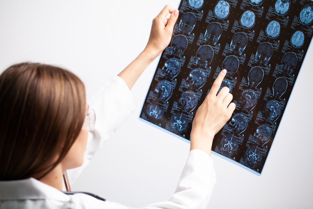
Diagnosis combines neurological examination with brain imaging studies.
The most commonly used techniques include:
- Brain magnetic resonance imaging (MRI).
- Computed tomography (CT).
- CT angiography.
- Magnetic resonance angiography.
- Vascular studies when necessary.
Magnetic resonance imaging generally provides better visualization of small infarctions located in deep brain structures.
Treatment
Treatment depends on the cause of the infarction and follows established protocols for ischemic stroke.
The goal is to preserve cerebral blood flow whenever possible, prevent recurrent cerebrovascular events, and promote functional recovery through rehabilitation programs tailored to each patient.
Management should always be individualized according to the clinical characteristics of each case.
Prognosis
The clinical course varies depending on several factors, including:
- The size of the infarction.
- The precise location of the lesion.
- The timeliness of diagnosis.
- The treatment received.
- The patient’s overall health status.
In many cases, functional recovery can be favorable, particularly when the lesion is small and rehabilitation is initiated early.
Conclusion
A basal ganglia infarction is a type of stroke that affects brain structures essential for the control of movement and other neurological functions. Its clinical manifestations depend on the region affected and may include motor, balance, and coordination impairments. Identifying this type of infarction through clinical assessment and neuroimaging is essential for understanding the characteristics of the lesion and guiding its management.
If you would like more information about NeuroAiD II, please complete this contact form.
"*" indicates required fields


