

Midbrain infarction is a rare type of ischemic stroke that affects the midbrain, the upper portion of the brainstem responsible for coordinating essential functions such as eye movements, posture, alertness, and various neural pathways connecting the brain with the rest of the body.
Although it accounts for only a small proportion of strokes, an infarction in this region can cause highly characteristic neurological symptoms, the severity of which depends on the precise location and extent of the lesion.
What is a midbrain infarction?
A midbrain infarction occurs when blood flow to this part of the brainstem is interrupted, preventing an adequate supply of oxygen and nutrients to neurons.
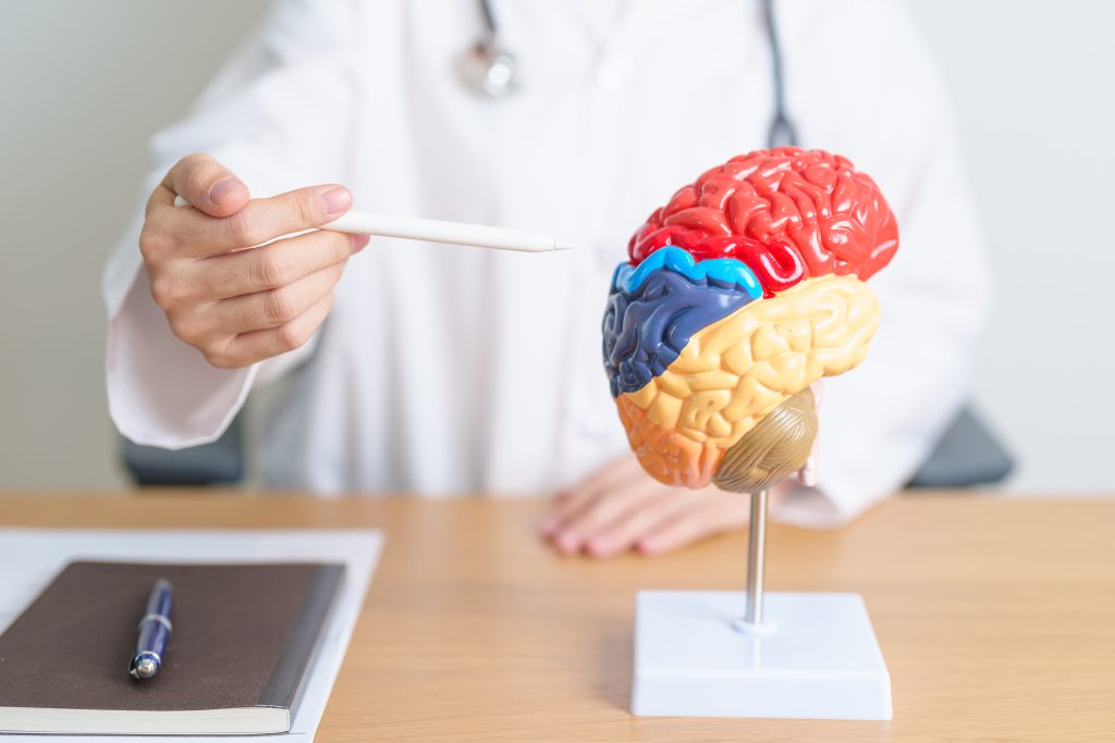
The midbrain contains important structures responsible for motor control, eye movements, coordination, and level of consciousness. Therefore, a lesion in this region can cause a combination of symptoms that helps localize the affected area of the brain.
What are the functions of the midbrain?
The midbrain plays a fundamental role in multiple neurological functions, including:
- Control of eye movements.
- Coordination of posture and balance.
- Regulation of muscle tone.
- Integration of visual and auditory reflexes.
- Involvement in alertness and consciousness.
- Transmission of information between the brain and the spinal cord.
Damage to any of these structures explains the wide range of clinical manifestations that may occur.
Causas
Most midbrain infarctions result from impaired vertebrobasilar circulation.
The most common causes include:
- Atherosclerosis.
- Arterial thrombosis.
- Cerebral embolism.
- Vertebral artery dissection.
- Small vessel disease.
- Coagulation disorders.
In some cases, the infarction may extend to other regions of the brainstem or be associated with lesions involving the thalamus or pons.
Symptoms of midbrain infarction
Symptoms depend on the affected vascular territory and the structures involved.
The most common symptoms include:
- Double vision (diplopia).
- Drooping of the eyelid (ptosis).
- Abnormal eye movements.
- Weakness on one side of the body.
- Coordination problems.
- Difficulty walking.
- Dizziness or vertigo.
- Balance disturbances.
- Changes in the level of consciousness.
- Difficulty speaking.
Some patients may also develop specific neurological syndromes, such as Weber, Benedikt, Claude, or Nothnagel syndrome, depending on the precise region of the midbrain affected.
How is it diagnosed?
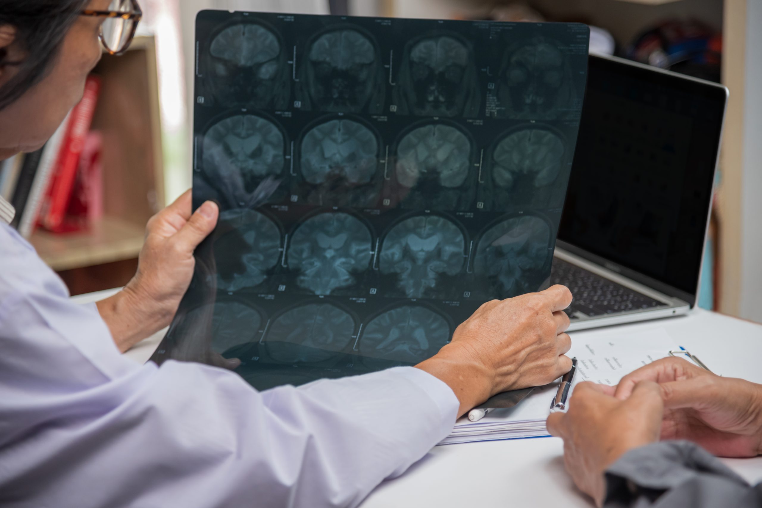
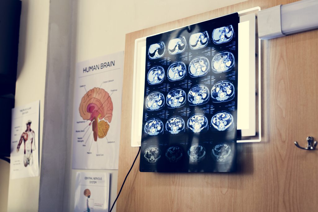
Diagnosis requires a clinical evaluation together with brain imaging studies.
The most commonly used diagnostic tests include:
- Brain magnetic resonance imaging (MRI).
- Computed tomography (CT).
- CT angiography (CTA).
- Magnetic resonance angiography (MRA).
- Vascular studies.
Magnetic resonance imaging is generally the most useful technique for detecting small lesions located in the brainstem.
Treatment
Treatment depends on the underlying cause of the infarction and follows established protocols for ischemic stroke.
The goal is to restore or preserve cerebral blood flow whenever possible, prevent further complications, and promote functional recovery through rehabilitation tailored to each patient’s individual needs.
Management should always be carried out under specialized medical supervision.
Prognosis
The outcome can vary considerably depending on:
- The size of the infarction.
- The speed of diagnosis.
- The region of the midbrain affected.
- The presence of other brain lesions.
- The patient’s overall condition.
Some patients experience significant functional recovery, while others may have persistent deficits affecting mobility, balance, or eye movements.
Conclusion
Midbrain infarction is a rare form of stroke that affects a key region of the brainstem. Due to the multiple functions performed by the midbrain, this type of lesion can produce a wide range of neurological symptoms. Identification through clinical examination and neuroimaging allows the lesion to be accurately localized and helps explain the neurological deficits experienced by each patient.
If you would like more information about NeuroAiD II, please complete this contact form.
"*" indicates required fields


