


A lobar hemorrhage is a type of intracerebral hemorrhage that occurs when a blood vessel ruptures within one of the lobes of the brain. Unlike other intracerebral hemorrhages that affect deeper brain structures, this type of bleeding occurs in areas responsible for language, memory, vision, movement, or behavior. As a result, its clinical manifestations can vary considerably depending on the region affected.
Although it accounts for a smaller proportion of hemorrhagic strokes, lobar hemorrhage requires urgent medical attention, as the bleeding can rapidly increase pressure within the skull and cause significant neurological damage.
What is a lobar hemorrhage?
A lobar hemorrhage is an accumulation of blood within the brain tissue located in one of the four lobes of the brain:
- Frontal lobe.
- Parietal lobe.
- Temporal lobe.
- Occipital lobe.
When a blood vessel ruptures, blood leaks into the surrounding brain tissue, compressing neurons and disrupting normal brain function.
The severity depends on the volume of bleeding, how rapidly it occurs, and the region of the brain affected.
What causes a lobar hemorrhage?
The causes are diverse and may vary depending on the patient’s age.
The most common include:
- High blood pressure.
- Cerebral amyloid angiopathy.
- Arteriovenous malformations.
- Aneurysms.
- Head trauma.
- Brain tumors associated with bleeding.
- Blood clotting disorders.
- Use of anticoagulant medications.
In older adults, one of the most common causes is cerebral amyloid angiopathy, a condition characterized by the accumulation of proteins in the walls of cerebral blood vessels, making them more fragile and prone to rupture.
What functions can be affected?
Each cerebral lobe performs different functions. Therefore, a lobar hemorrhage does not always cause the same symptoms.
Frontal lobe
It may cause:
- Weakness on one side of the body.
- Personality changes.
- Difficulty planning tasks.
- Behavioral changes.
- Speech difficulties when the dominant hemisphere is affected.
Parietal lobe
Common symptoms include:
- Loss of sensation.
- Difficulty recognizing objects by touch.
- Problems with spatial orientation.
- Difficulty performing coordinated movements.
Temporal lobe
Symptoms may include:
- Memory impairment.
- Difficulty understanding language.
- Seizures.
- Emotional changes.
Occipital lobe
Symptoms are usually related to vision:
- Partial loss of the visual field.
- Blurred vision.
- Difficulty recognizing images.
Symptoms of a lobar hemorrhage
The onset is usually sudden.
The most common symptoms include:
- Severe headache.
- Weakness on one side of the body.
- Difficulty speaking.
- Visual disturbances.
- Confusion.
- Drowsiness.
- Nausea and vomiting.
- Seizures.
- Loss of consciousness in more severe cases.
Symptoms depend on both the size of the hematoma and the region of the brain affected.
How is it diagnosed?
When a cerebral hemorrhage is suspected, rapid diagnosis is essential.
The most commonly used tests include:
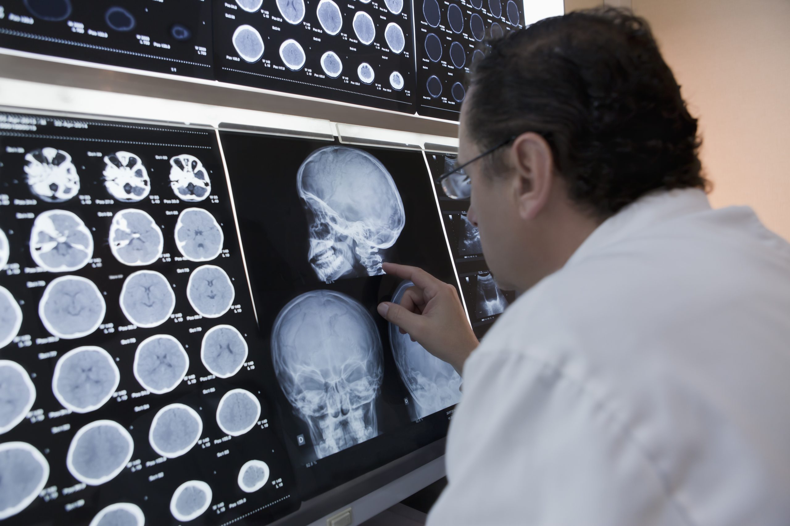
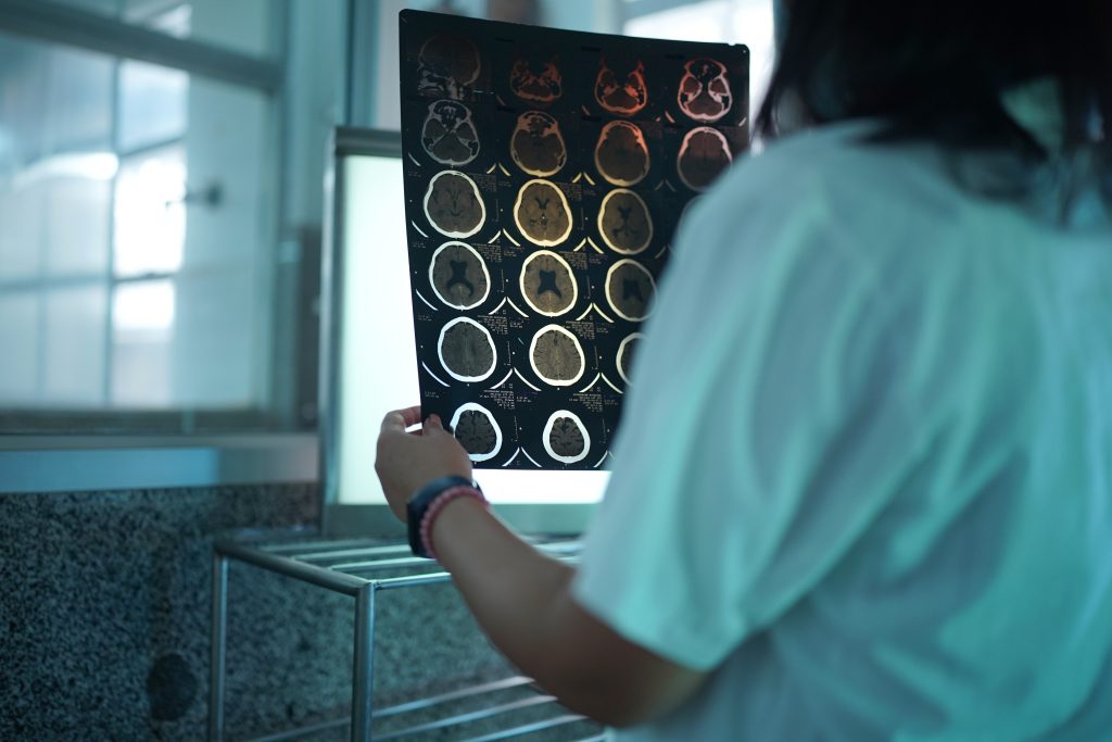
- Computed tomography (CT), which can quickly confirm the presence of bleeding.
- Magnetic resonance imaging (MRI), which provides a more detailed assessment of the lesion and can help identify possible causes.
- CT angiography (CTA) or cerebral angiography when vascular malformations or aneurysms are suspected.
Blood tests are also commonly performed to assess coagulation and identify other possible abnormalities.
Treatment

Treatment depends on several factors:
- Size of the hematoma.
- Location.
- The patient’s neurological status.
- Age.
- Cause of the bleeding.
Initial treatment focuses on stabilizing the patient and preventing further bleeding.
This may include:
- Strict blood pressure control.
- Correction of coagulation abnormalities.
- Treatment to reduce intracranial pressure when necessary.
- Monitoring in specialized units.
In certain cases, surgery may be required to evacuate the hematoma or treat the underlying cause of the bleeding.
Possible complications
Complications may occur both during the acute phase and throughout recovery.
These may include:
- Cerebral edema.
- Seizures.
- Increased intracranial pressure.
- Cognitive difficulties.
- Language impairments.
- Motor impairments.
- Difficulty walking.
- Visual deficits.
The risk depends primarily on the size of the hemorrhage and how quickly medical attention is received.
Recovery
Each patient’s recovery is different.
In many cases, rehabilitation begins within a few days after the patient has been medically stabilized.
It may include:
- Physiotherapy to restore strength and mobility.
- Occupational therapy to promote independence in daily activities.
- Speech and language therapy when there are difficulties with speech or swallowing.
- Neuropsychological rehabilitation to address memory, attention, and executive functions.
Recovery is often most pronounced during the first few months, although some people may continue to regain function over a much longer period.
Can it be prevented?
Not all lobar hemorrhages can be prevented, but the risk can be reduced by managing modifiable risk factors.
The main recommendations include:
- Maintaining healthy blood pressure.
- Avoiding smoking.
- Limiting alcohol consumption.
- Following prescribed anticoagulant therapy correctly when indicated.
- Attending regular medical check-ups.
- Managing diabetes and cholesterol levels.
Conclusion
A lobar hemorrhage is a type of cerebral hemorrhage that directly affects one of the lobes of the brain, meaning its clinical manifestations can vary widely depending on the region affected. Early recognition of symptoms, rapid diagnosis, and appropriate treatment are essential for improving prognosis. After the acute phase, rehabilitation plays a key role in restoring function and promoting the greatest possible level of independence.
If you would like more information about NeuroAiD II, please complete this contact form.
"*" indicates required fields


