



Weber syndrome is a rare neurological syndrome that occurs when a lesion affects the ventral portion of the midbrain, a region of the brainstem responsible for transmitting motor signals between the brain and the rest of the body. Its most characteristic feature is the combination of third cranial nerve palsy on the same side as the lesion and weakness on the opposite side of the body.
What is Weber syndrome?

Weber syndrome is an alternating midbrain syndrome caused by a lesion that simultaneously affects:
- The oculomotor nerve (third cranial nerve).
- The corticospinal tract.
- In some cases, the corticonuclear tract.
This combination results in ocular abnormalities on one side and motor deficits on the opposite side of the body.
Where is the lesion located?
The lesion is located in the ventral portion of the midbrain, specifically in the region of the cerebral peduncles.
This area contains numerous nerve fibers involved in:
- Voluntary movement.
- Facial movement.
- Control of the eye muscles.
- Transmission of information between the cerebral cortex and the spinal cord.
A lesion in this area produces a highly characteristic neurological presentation.
What is the most common cause?
The most common cause is an ischemic stroke affecting small branches of the posterior cerebral artery.
Other less common causes include:
- Midbrain hemorrhage.
- Tumors.
- Multiple sclerosis.
- Vascular malformations.
- Traumatic brain injury.
The sudden onset of symptoms usually suggests a vascular origin.
Symptoms of Weber syndrome
The onset is usually sudden when the underlying cause is a cerebral infarction.
Third cranial nerve palsy
Involvement of the oculomotor nerve causes symptoms on the same side as the lesion:
- Drooping of the eyelid (ptosis).
- Difficulty moving the eye.
- Double vision.
- Dilated pupil.
- Downward and outward deviation of the eye.
These signs result from the loss of control of several extraocular muscles.
Weakness on the opposite side of the body
Damage to the corticospinal tract can cause:
- Hemiparesis.
- Hemiplegia.
- Difficulty walking.
- Loss of strength in the contralateral arm and leg.
When the corticonuclear fibers are also affected, facial movement abnormalities may occur.
Other possible symptoms
Depending on the extent of the lesion, additional symptoms may include:
- Dysarthria.
- Motor clumsiness.
- Slowness of movement.
- Gait disturbances.
- Muscle fatigue.
In some patients, other neurological deficits may coexist if the infarction extends into adjacent regions of the midbrain.
How is it diagnosed?
Diagnosis is based on neurological examination and neuroimaging studies.
The combination of:
- Ipsilateral third cranial nerve palsy.
- Contralateral hemiplegia.
is highly suggestive of Weber syndrome.
Imaging studies commonly used include:
- Brain magnetic resonance imaging.
- Diffusion-weighted magnetic resonance imaging to detect acute infarction.
- Magnetic resonance angiography.
- Computed tomography angiography.
- Computed tomography during the initial assessment.
In addition, cardiovascular and vascular investigations are usually performed to determine the underlying cause of the stroke.
Differences from other midbrain syndromes
Several syndromes affect the midbrain and share certain clinical features, although important differences exist between them.
Claude syndrome
The predominant features include:
- Third cranial nerve palsy.
- Contralateral ataxia.
- Coordination deficits.
Benedikt syndrome
It is characterized by:
- Ocular palsy.
- Tremor.
- Involuntary movements.
- Ataxia.
Nothnagel syndrome
It mainly causes:
- Cerebellar abnormalities.
- Balance disturbances.
- Eye movement disorders.
The presence of marked hemiplegia distinguishes Weber syndrome from these other midbrain syndromes.
Clinical course and prognosis
Recovery depends on several factors, including:
- The size of the infarction.
- How quickly treatment is initiated.
- The patient’s age.
- The presence of vascular risk factors.
Some patients may partially recover ocular mobility and muscle strength over the following months, while others may experience permanent neurological deficits.
Rehabilitation

Rehabilitation is usually multidisciplinary and includes different interventions according to each patient’s individual needs.
These may include:
- Physical therapy to restore strength and improve gait.
- Occupational therapy to improve independence in daily activities.
- Visual rehabilitation when eye movement abnormalities persist.
- Speech and language therapy when difficulties with speech or swallowing are present.
The intensity of rehabilitation depends on the severity of the neurological impairment.
Conclusion
Weber syndrome is a neurological syndrome typically caused by an infarction involving the ventral portion of the midbrain. The combination of third cranial nerve palsy on one side and hemiplegia on the opposite side of the body is its main clinical feature. Recognition of this pattern allows rapid localization of the lesion and helps identify an alternating brainstem syndrome, facilitating early neurological assessment and the initiation of appropriate treatment and rehabilitation.
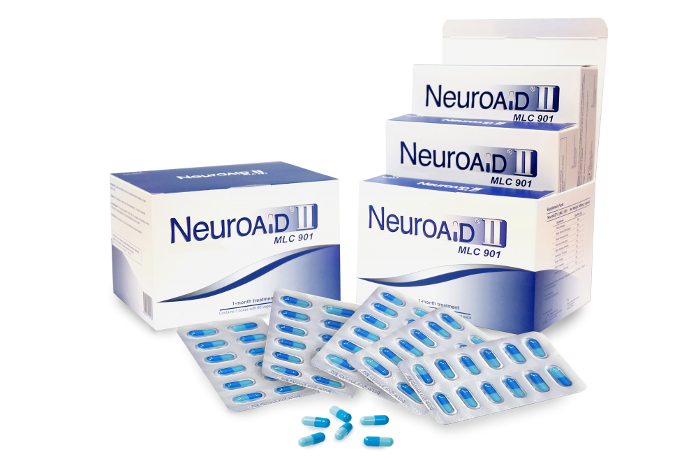
If you need information about NeuroAiD II, please complete this contact form
"*" indicates required fields


