

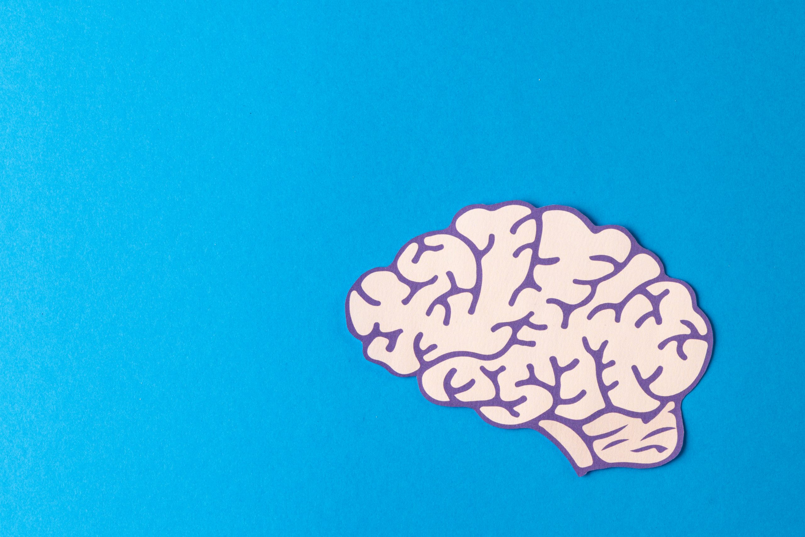

Cerebral white matter is an essential part of the central nervous system that enables communication between different regions of the brain. Although it often receives less attention than gray matter, it plays a fundamental role in the rapid transmission of information required for movement, memory, language, thinking, and other cognitive functions.
White matter abnormalities can occur as a result of aging, vascular diseases, trauma, demyelinating disorders, or stroke. Depending on the extent and location of the injury, clinical manifestations can range from mild changes to significant neurological impairment.
What is cerebral white matter?
White matter is primarily composed of millions of nerve fibers called axons, which connect neurons to one another. These fibers are covered by myelin, a lipid-rich substance that acts as an insulator and allows nerve impulses to travel rapidly and efficiently.
Its whitish appearance is due precisely to the abundance of myelin, which distinguishes it from gray matter, composed mainly of neuronal cell bodies.
Where is it located?
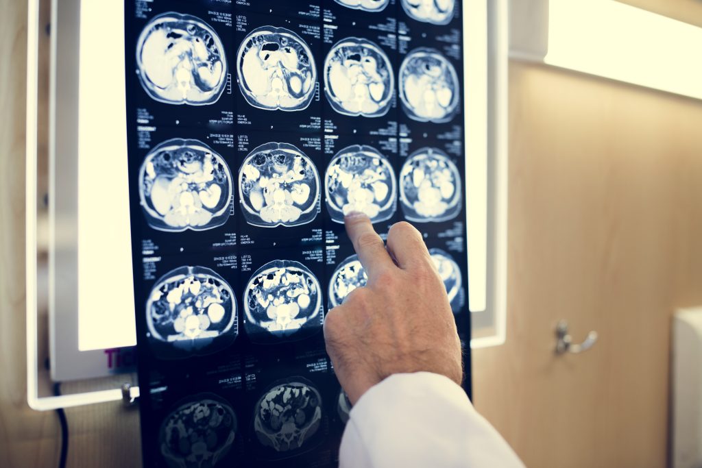
White matter occupies a large portion of the interior of the cerebral hemispheres and surrounds various deep brain structures.
Its main components include:
- Corpus callosum.
- Internal capsule.
- Corona radiata.
- Association fibers.
- Commissural fibers.
- Projection fibers.
These pathways allow different regions of the brain to work together in a coordinated manner.
What is its function?
The primary function of white matter is to transmit information between different areas of the nervous system.
It makes it possible to:
- Coordinate movement.
- Integrate sensory information.
- Process language.
- Support memory.
- Facilitate learning.
- Coordinate complex cognitive functions.
- Enable communication between the two cerebral hemispheres.
Without these connections, different areas of the brain would function in isolation.
Difference between white matter and gray matter
Although they work together, these two structures perform different functions.
Gray matter:
- Processes information.
- Contains neuronal cell bodies.
- Is located primarily in the cerebral cortex and certain deep brain nuclei.
White matter:
- Transmits information between different regions.
- Is composed primarily of myelinated axons.
- Acts as an internal communication network within the brain.
Both are essential for the normal functioning of the nervous system.
What diseases can affect white matter?
Several conditions can cause damage to cerebral white matter.
The most common include:
- Ischemic stroke.
- Cerebral hemorrhage.
- Small vessel disease.
- Multiple sclerosis.
- Traumatic brain injury.
- Leukoaraiosis.
- Neurodegenerative diseases.
- Central nervous system infections.
- Brain tumors.
The underlying cause will determine both the symptoms and the appropriate treatment.
White matter and stroke
In stroke, lesions can directly affect the white matter when blood flow to the deep nerve fibers is interrupted.
Some particularly vulnerable structures include:
- Internal capsule.
- Corona radiata.
- Centrum semiovale.
When these pathways are damaged, significant motor, sensory, or cognitive impairments may occur.
Symptoms of a white matter injury
Symptoms depend on the location and extent of the damage.
The most common include:
- Weakness on one side of the body.
- Loss of sensation.
- Balance problems.
- Slower walking.
- Language impairments.
- Memory difficulties.
- Reduced processing speed.
- Attention difficulties.
- Mood changes.
- Urinary incontinence in some cases.
In many older adults, the accumulation of small white matter lesions may contribute to progressive cognitive decline.
It is relatively common for magnetic resonance imaging (MRI) to detect small white matter abnormalities in older adults.
These lesions are often associated with:
- High blood pressure.
- Diabetes.
- High cholesterol.
- Small vessel disease.
- Brain aging.
In many cases, they do not cause significant symptoms, although when extensive, they may affect gait, memory, or mental processing speed.
How are white matter lesions diagnosed?
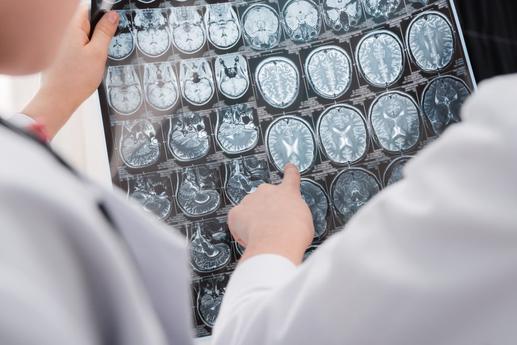
Diagnosis combines clinical assessment with various imaging tests.
The main tests include:
- Magnetic resonance imaging (MRI), considered the most sensitive imaging technique.
- Computed tomography (CT).
- Vascular imaging studies.
- Neuropsychological assessment when cognitive impairments are present.
MRI provides detailed visualization of the location and extent of white matter lesions.
Conclusion
Cerebral white matter forms the communication network that connects different regions of the brain and enables the nervous system to function properly. It plays an essential role in coordinating movement, integrating sensory information, processing language, and supporting complex cognitive functions.
When white matter is damaged by a stroke, vascular disease, or another medical condition, motor, sensory, or cognitive impairments of varying severity may occur. Early diagnosis, treatment of the underlying cause, and a rehabilitation program tailored to each patient are essential for promoting functional recovery and preserving quality of life.
If you would like more information about NeuroAiD II, please complete this contact form.
"*" indicates required fields


