

A medial pontine infarction is a type of ischemic stroke that affects the central portion of the pons, a structure located within the brainstem that is responsible for transmitting information between the brain, cerebellum, and spinal cord. Although it accounts for a small proportion of strokes, it can cause significant neurological deficits due to the large number of neural pathways that pass through this region.
Most medial pontine infarctions are caused by occlusion of small perforating arteries arising from the basilar artery. Depending on the size and location of the lesion, symptoms can range from mild weakness to complex neurological syndromes affecting movement, sensation, and eye movement control.
What functions does the pons perform?

The pons is part of the brainstem and plays an essential role in communication between different regions of the nervous system.
Its main functions include:
- Transmitting signals between the cerebral cortex and the spinal cord.
- Contributing to motor coordination in conjunction with the cerebellum.
- Controlling certain eye movements.
- Regulating functions related to breathing.
- Contributing to alertness and consciousness.
- Containing the nuclei of several cranial nerves.
The medial region of the pons contains numerous motor and sensory fibers, which means that a lesion in this area can affect multiple functions simultaneously.
What causes a medial pontine infarction?
The most common cause is occlusion of the paramedian perforating arteries that supply the medial portion of the pons.
The main risk factors include:
- Hypertension.
- Diabetes mellitus.
- High cholesterol.
- Smoking.
- Small vessel disease.
- Atherosclerosis of the basilar artery.
- Advanced age.
In some patients, the infarction may be associated with large-artery atherosclerotic disease or, less commonly, a cardioembolic source.
Symptoms of a medial pontine infarction
Clinical manifestations depend on the extent of brain damage and the specific structures affected.
The most common symptoms include:
Weakness on one side of the body
The weakness typically affects both the arm and the leg and may impair walking and the ability to perform activities of daily living.
Sensory disturbances
If the ascending sensory pathways are affected, the following symptoms may occur:
- Reduced sense of touch.
- Impaired vibration sensation.
- Loss of proprioception.
- Numbness.
These sensory disturbances typically occur on the side of the body opposite to the infarction.
Eye movement disorders
When the infarction involves the fibers of certain cranial nerves, the following symptoms may occur:
- Double vision.
- Difficulty moving one eye.
- Gaze deviation.
- Impaired control of eye movements.
In some cases, these lesions may be associated with specific brainstem syndromes, such as Raymond syndrome, Foville syndrome, or Millard-Gubler syndrome, depending on the structures involved.
Coordination impairments
Some patients may experience:
- Unsteadiness while walking.
- Clumsiness of movement.
- Difficulty maintaining balance.
These symptoms may occur when connections with the cerebellum are also affected.
Other symptoms
Depending on the size of the infarction, other symptoms may also occur:
- Difficulty speaking (dysarthria).
- Difficulty swallowing (dysphagia).
- Dizziness.
- Nausea.
- Altered level of consciousness in extensive lesions.
How is it diagnosed?

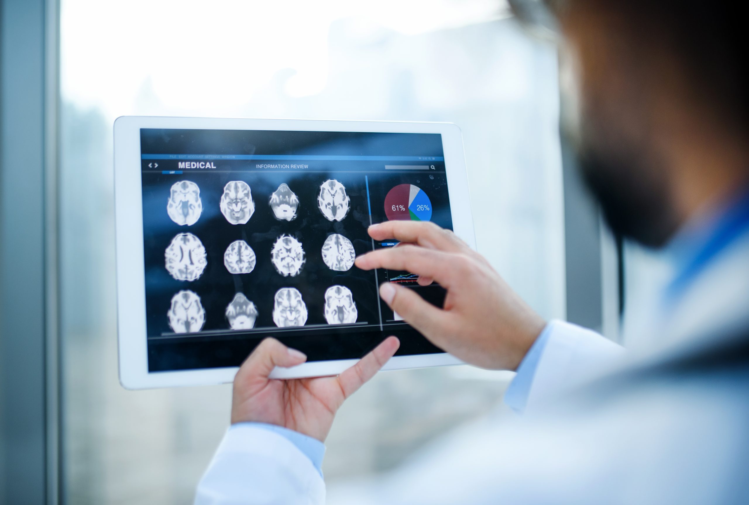
Diagnosis begins with a detailed neurological examination, followed by neuroimaging studies to confirm the location of the infarction.
The most commonly used techniques include:
- Brain magnetic resonance imaging with diffusion-weighted imaging.
- Computed tomography.
- Computed tomography angiography or magnetic resonance angiography.
- Intracranial vascular imaging studies.
- Cardiac evaluation when an embolic source is suspected.
Magnetic resonance imaging is particularly useful for detecting small brainstem infarctions that may not be visible on an initial computed tomography scan.
Treatment
Treatment follows the general recommendations for ischemic stroke and depends on how quickly the patient receives medical attention.
During the acute phase, the primary goal is to restore blood flow when possible and prevent complications.
Subsequent management typically includes:
- Blood pressure control.
- Management of diabetes and high cholesterol.
- Prevention of recurrent stroke.
- Intensive physical rehabilitation.
- Occupational therapy.
- Speech and swallowing rehabilitation when dysphagia or dysarthria is present.
The treatment plan should be tailored to each patient’s specific neurological deficits and recovery needs.
Prognosis
Recovery depends on the size of the infarction, the structures affected, and how quickly treatment is initiated.
Small infarctions may have a favorable outcome, with substantial functional recovery supported by rehabilitation. In contrast, more extensive pontine lesions may result in persistent motor deficits and may even compromise vital functions when critical areas of the brainstem are affected.
Early rehabilitation plays a key role in promoting the recovery of mobility, coordination, and functional independence.
The importance of medial pontine infarction
Medial pontine infarction highlights the critical role of the brainstem in the functioning of the nervous system. A localized lesion in this region can impair movement, sensation, eye movements, and other functions essential for daily living.
Recognizing symptoms consistent with a pontine stroke enables early diagnosis and the prompt initiation of treatment and rehabilitation, which are key factors in improving prognosis and reducing the risk of permanent neurological deficits.
If you would like more information about NeuroAiD II, please complete this contact form.
"*" indicates required fields


