


Occipital stroke and visual disturbances are closely related because the occipital lobe is the region of the brain responsible for processing visual information. When a stroke occurs in this area, the ability to correctly interpret visual signals received from the eyes can be significantly impaired. Although the eyes themselves may remain structurally and functionally intact, the brain may partially or completely lose its ability to process visual information.
Understanding the relationship between occipital stroke and visual disturbances is essential for recognizing early symptoms, ensuring timely medical intervention, and improving the chances of recovery. Many people are unaware that sudden vision loss can be one of the primary warning signs of a stroke.
What is an occipital stroke?
An occipital stroke is a cerebrovascular event that affects the occipital lobe, located at the back of the brain. This region is responsible for receiving and processing visual information transmitted from the eyes.
When blood flow to this area is disrupted by a blood clot or hemorrhage, neurons involved in visual processing may sustain damage. As a result, visual disturbances may develop, representing one of the most characteristic clinical manifestations of an occipital stroke.
The nature and severity of visual symptoms can vary considerably depending on the extent and precise location of the brain lesion.
Why do visual disturbances occur after an occipital stroke?
The relationship between occipital stroke and visual disturbances arises from the brain’s role in interpreting visual information received from the eyes.
The eyes may function normally and transmit visual signals without impairment. However, when the occipital lobe is damaged, the brain may be unable to process these signals correctly.
This means that an individual may have normal ophthalmological examination findings while still experiencing significant visual impairments following an occipital stroke.
Main visual disturbances associated with an occipital stroke

Visual disturbances associated with an occipital stroke can manifest in various ways.
Homonymous hemianopia
It is one of the most common visual disturbances associated with an occipital stroke.
Individuals with homonymous hemianopia experience loss of vision in either the right or left half of the visual field in both eyes.
For example, a person may be unable to see objects located on the left side of their surroundings, even though their eyes remain structurally healthy.
Homonymous hemianopia can interfere with everyday activities such as:
-
Reading.
-
Driving.
-
Walking.
-
Recognizing objects.
In the context of occipital stroke and visual disturbances, homonymous hemianopia is one of the most characteristic clinical manifestations.
Quadrantanopia
When the damage affects only a specific region of the visual cortex, vision loss may be limited to a single quadrant of the visual field.
In these cases, individuals lose the ability to perceive visual information within a specific quadrant of their field of vision.
Quadrantanopia is generally less disabling than complete homonymous hemianopia, but it can still significantly affect daily functioning and quality of life.
Cortical blindness
Another significant visual impairment associated with occipital stroke is cortical blindness.
This condition occurs when both occipital lobes sustain extensive damage, resulting in the brain’s inability to process and interpret visual information.
Although the eyes may remain structurally and functionally intact, the individual may experience complete loss of conscious visual perception, resulting in functional blindness.
Visual agnosia
Some individuals may retain the ability to see objects but be unable to recognize or identify them.
This neurological condition is known as visual agnosia.
For example, a patient may be able to see a cup but remain unable to identify or recognize the object.
Visual agnosia illustrates how visual disturbances associated with an occipital stroke do not always involve vision loss but may also include impairments in the brain’s ability to interpret and recognize visual information.
Visual hallucinations
In some cases, individuals may perceive images, lights, or shapes that are not actually present.
These visual hallucinations can result from disruptions in the neural circuits responsible for processing visual information.
Although they may cause concern, visual hallucinations are among the neurological manifestations that can occur following a stroke.
Symptoms that may accompany visual disturbances
In addition to visual impairments, an occipital stroke may be accompanied by other neurological symptoms.
These may include:
-
Dizziness.
-
Sudden-onset headache.
-
Balance disturbances.
-
Difficulties with spatial orientation.
-
Confusion.
-
Visual memory impairments.
The combination of sudden vision loss and any of these neurological symptoms should be considered a medical emergency requiring immediate medical attention.
How is an occipital stroke diagnosed?
The diagnosis of an occipital stroke and associated visual disturbances requires a comprehensive neurological assessment.
The physician may perform the following examinations:
-
Neurological examination.
-
Visual field assessment.
-
Computed tomography (CT) scan.
-
Brain magnetic resonance imaging (MRI).
-
Cerebrovascular imaging studies.
Neuroimaging techniques help identify the precise location of the stroke and confirm involvement of the occipital lobe.
Treatment of occipital stroke
Treatment depends on whether the stroke is ischemic or hemorrhagic.
In cases of ischemic stroke, treatment options may include:
-
Intravenous thrombolytic therapy.
-
Mechanical thrombectomy.
-
Antiplatelet medications.
For hemorrhagic stroke, management may involve:
-
Blood pressure control.
-
Neurosurgical intervention.
-
Specialized neurological intensive care.
Timely medical intervention is essential to minimize brain damage and reduce the severity of associated visual impairments.
Visual recovery after an occipital stroke
The progression of visual impairments following an occipital stroke varies considerably between patients.
Some individuals experience spontaneous improvements during the first few months following a stroke, supported by the brain’s neuroplasticity mechanisms.
However, others may experience persistent or permanent visual deficits.
Visual rehabilitation may include:
-
Visual field training.
-
Occupational therapy.
-
Visual scanning exercises.
-
Environmental adaptations.
-
Use of specialized optical aids.
Early rehabilitation intervention is generally associated with better functional outcomes.
Impact on daily life

Occipital stroke and associated visual disturbances can significantly affect various activities of daily living.
The most commonly affected activities include:
-
Reading.
-
Using electronic devices.
-
Driving.
-
Independent mobility.
-
Facial recognition.
-
Work-related activities.
Many individuals develop compensatory strategies to adapt to their visual impairments and regain a degree of functional independence.
Risk factors for occipital stroke
The risk factors for occipital stroke are similar to those associated with other types of stroke:
-
Arterial hypertension.
-
Diabetes mellitus.
-
Hypercholesterolemia.
-
Smoking.
-
Obesity.
-
Physical inactivity.
-
Atrial fibrillation.
-
Cardiovascular disease.
Effective management of these risk factors can significantly reduce the likelihood of experiencing a stroke.
Conclusion
The relationship between occipital stroke and visual disturbances is one of the most characteristic clinical features of cerebrovascular events affecting the occipital lobe. Partial visual field loss, cortical blindness, visual agnosia, and difficulties interpreting visual information may occur even when the eyes remain structurally and functionally intact.
Early recognition of symptoms associated with occipital stroke and visual disturbances is essential for ensuring timely medical intervention and improving the chances of neurological recovery, ultimately contributing to better quality of life and greater functional independence for patients.
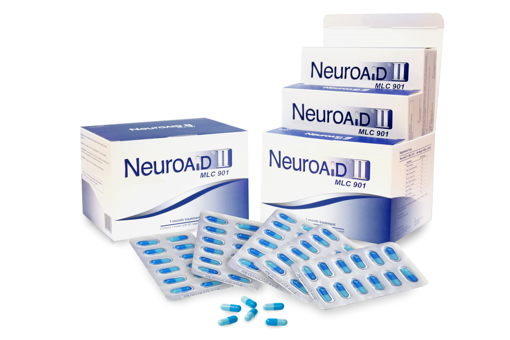
If you would like more information about NeuroAiD II, please complete this contact form.
"*" indicates required fields


