


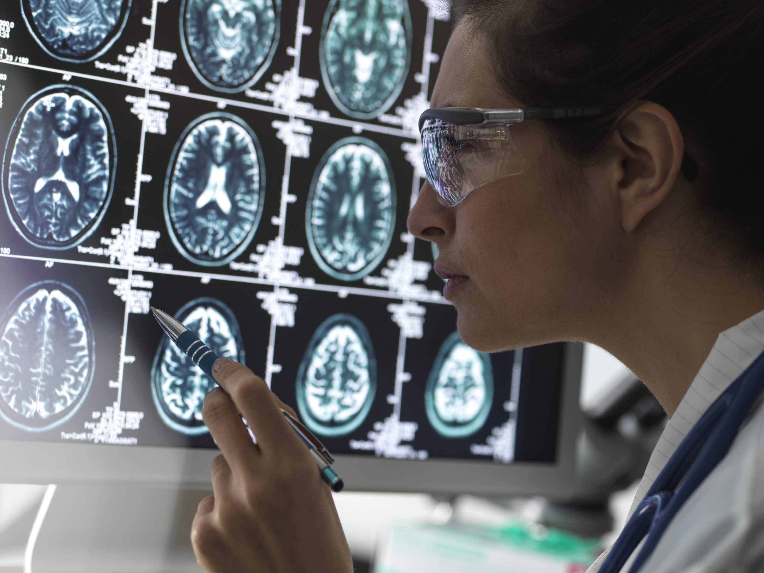

Raymond syndrome is a rare neurological syndrome caused by a lesion in the medial pons, a structure of the brainstem that connects different regions of the brain with the spinal cord. The most common cause is ischemic stroke, although it may also occur as a result of hemorrhage, tumors, or inflammatory diseases.

This syndrome is characterized by a combination of sixth cranial nerve (abducens nerve) palsy on the same side as the lesion and weakness on the opposite side of the body, a pattern that helps neurologists accurately localize the affected area.
Which structures are affected?
The lesion is located in the medial region of the pons, where several important neural pathways pass.
The main structures affected include:
- Abducens nerve fascicle.
- Corticospinal tract.
- Motor pathways connecting the brain to the spinal cord.
The simultaneous involvement of these nerve fibers explains why ocular symptoms and motor deficits can occur at the same time.
Symptoms of Raymond syndrome
Symptoms may vary depending on the size of the infarction or lesion, although the most common include:
- Inability to move one eye outward.
- Double vision (diplopia).
- Strabismus due to inward deviation of the eye.
- Weakness or paralysis on the opposite side of the body.
- Difficulty walking.
- Reduced dexterity in one hand or leg.
In some patients, motor impairment may be mild if the corticospinal fibers are only partially affected.
What causes Raymond syndrome?
The most common cause is a pontine infarction resulting from occlusion of small arteries supplying the brainstem.
Other possible causes include:
- Intracerebral hemorrhage.
- Multiple sclerosis.
- Brainstem tumors.
- Vascular malformations.
- Inflammatory or infectious processes.
Identifying the underlying cause of the lesion is essential for guiding treatment.
What causes Raymond syndrome?
The most common cause is a pontine infarction resulting from occlusion of small arteries supplying the brainstem.
Other possible causes include:
- Intracerebral hemorrhage.
- Multiple sclerosis.
- Brainstem tumors.
- Vascular malformations.
- Inflammatory or infectious processes.
Identifying the underlying cause of the lesion is essential for guiding treatment.
How is Raymond syndrome diagnosed?
Diagnosis begins with a detailed neurological examination, with particular attention to eye movements and muscle strength.
The most commonly used diagnostic tests include:
- Brain magnetic resonance imaging (MRI).
- Computed tomography (CT).
- CT angiography (CTA) or MR angiography (MRA) when a vascular abnormality is suspected.
- Comprehensive neurological examination.
MRI can usually confirm the precise location of the lesion within the pons.
Treatment
Treatment depends on the underlying condition responsible for the syndrome.
When the cause is a stroke, management includes established stroke treatment protocols, together with control of cardiovascular risk factors.
Subsequently, rehabilitation plays an important role in improving mobility and ocular function. Depending on the residual deficits, the rehabilitation program may involve physical therapists, occupational therapists, and neurological rehabilitation specialists.
Prognosis

The clinical course can vary considerably. In patients with small lesions, ocular motility and muscle strength may improve significantly during the months following the injury.
In other cases, residual neurological deficits may persist, particularly when the damage involves a larger area of the brainstem or when treatment is delayed.
The importance of Raymond syndrome
Although it is a rare condition, Raymond syndrome is a classic example of how a highly localized lesion can produce a characteristic pattern of neurological symptoms. Recognizing the association between sixth cranial nerve palsy and weakness on the opposite side of the body can help rapidly localize the lesion to the pons, facilitating early diagnosis and appropriate patient management.
If you need information about NeuroAiD II, please complete this contact form
"*" indicates required fields


